Dental X-rays: when they are used and what an image may show
A dental X-ray can provide additional information about teeth and nearby tissues when a visual examination alone is not enough. A dentist decides whether an image is appropriate based on the examination, symptoms, health history, and any previous images available.
What is a dental X-ray?
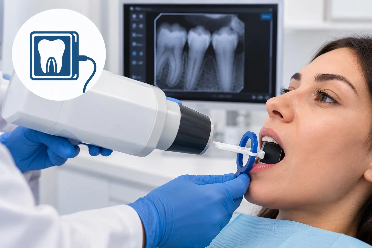
It is an image made using X-rays to help assess structures inside the mouth. For an intraoral digital image, a small sensor is placed inside the mouth and the image is recorded electronically.
When might one be recommended?
An image may be suggested when the dentist needs more information about a finding from the examination, wants to assess a tooth root or surrounding tissues, or is planning care. The decision is individual; an X-ray is not needed at every visit.
What can an image show?
Depending on the type of image and the area covered, an X-ray may help assess parts of a tooth that are not directly visible, its root, and nearby bone, and may reveal some changes that cannot be seen during a visual examination. The image is considered together with the examination and other information; it is not a complete diagnosis by itself.
What happens during the procedure?
The dentist explains the procedure and places a small sensor inside the mouth near the area being imaged. You will be asked to hold still briefly while the image is taken. If the position feels uncomfortable, tell the dentist so it can be adjusted.
What equipment is used at the clinic?
Dental Clinic Kapital 2022 uses a digital intraoral dental imaging sensor (viziograph). The image is displayed on an all-in-one computer, where the dentist can review it promptly and consider it when planning care.
Price at Dental Clinic Kapital 2022
Price — 1,000 AMD